Radius Bone Labelled Diagram : Similar Images, Stock Photos & Vectors of Osteoporosis ... - In its distal part, the radial shaft expands to form a rectangular end.
byAdmin•
0
Radius Bone Labelled Diagram : Similar Images, Stock Photos & Vectors of Osteoporosis ... - In its distal part, the radial shaft expands to form a rectangular end.. The radius bone is this bone here and it lies laterally in the anatomical position. Human body bone joint pains anatomy humerus with radius and ulna bones. Related posts of labelled diagram of radius bone related posts of labelled diagram of radius bone. It's not that clear on this model here, but i'll switch over to another diagram and show you. The tendon of the brachioradialis.
Learn everything about the anatomy of radius and ulna with our articles, video tutorials, labeled diagrams, and quizzes. Axial and appendicular system review lab anatomy & physiology 201 with radius anatomy pictures and information these pictures of this page are about:radius bone location. This ulnar view labelled illustration is from 'asklepios atlas of the human anatomy'. The radius or radial bone is one of the two large bones of the forearm, the other being the ulna. These are all the terms from all.
Wrist Bones Diagram — UNTPIKAPPS from www.untpikapps.com The radius bone is a long horizontal bone present in the forearm and is also called the radial bone. Projection of bone on the lateral surface of the distal radius bone. The radius bone is shorter. Skull, clavicle, mandible, scapula, thorax, sternum, humerus, ulna, radius, carpus, phalanges (fingers), metacarpus, spine, pelvis, sacrum, femur, tibia, fibula, tarsus. Radius, in anatomy, the outer of the two bones of the forearm when viewed with the palm facing forward. The skeleton acts as a scaffold by providing support and protection for the soft tissues that make up the rest of the body. Radius bone is a photograph by asklepios medical atlas which was uploaded on august 3rd, 2016. Learn everything about the anatomy of radius and ulna with our articles, video tutorials, labeled diagrams, and quizzes.
The tendon of the brachioradialis.
I'm not sure of what you mean by bone diagram. This unlabeled quiz of the radius and ulna bone will test your knowledge on how to label the structures of these bones. The radius bone is this bone here and it lies laterally in the anatomical position. Its upper concave surface articulates with the. It extends obliquely downward into a strong, conical projection. Proximal radius (head, neck and tuberosity). Related posts of labelled diagram of radius bone related posts of labelled diagram of radius bone. The photograph may be purchased as wall art, home decor, apparel, phone cases, greeting cards, and more. It extends from the lateral side of the elbow to the thumb side of the wrist and runs parallel to the ulna. Learn vocabulary, terms and more with flashcards, games and other study tools. The ulna articulates with the trochlea and the radius articulates with the capitulum. Radius bone is a photograph by asklepios medical atlas which was uploaded on august 3rd, 2016. A basic human skeleton is studied in schools with a simple diagram.
The photograph may be purchased as wall art, home decor, apparel, phone cases, greeting cards, and more. Related posts of labelled diagram of radius bone related posts of labelled diagram of radius bone. All land vertebrates have this bone. The radius or radial bone is one of the two large bones of the forearm, the other being the ulna. Learn vocabulary, terms and more with flashcards, games and other study tools.
Bone Anatomy Labeled Diagram Stock Vector - Illustration ... from thumbs.dreamstime.com The radius is a long bone in the forearm. 12 photos of the labelled diagram of radius bone. 769 x 1000 jpeg 102kb. This ulnar view labelled illustration is from 'asklepios atlas of the human anatomy'. Skull, clavicle, mandible, scapula, thorax, sternum, humerus, ulna, radius, carpus, phalanges (fingers), metacarpus, spine, pelvis, sacrum, femur, tibia, fibula, tarsus. Labeled radius anterior view left radius coxal bone unlabeled long bone diagram unlabeled radius bone landmarks radius radial tuberosity neck of radius bone radius vs ulna bone ulna bone location radius and ulna atlas radius bone features tibia fibula radius ulna where is the. Radius, in anatomy, the outer of the two bones of the forearm when viewed with the palm facing forward. The radius or radial bone is one of the two large bones of the forearm, the other being the ulna.
All land vertebrates have this bone.
Short video describing the skeletal structures of the radiusstructures identified:headneckradial tuberositystyloid process of the radiusulnar notch. Its upper concave surface articulates with the. The bones mentioned in each human skeleton chart are: Lower end of radius bone. Learn vocabulary, terms and more with flashcards, games and other study tools. In its distal part, the radial shaft expands to form a rectangular end. Related posts of labelled diagram of radius bone related posts of labelled diagram of radius bone. Axial and appendicular system review lab anatomy & physiology 201 with radius anatomy pictures and information these pictures of this page are about:radius bone location. Labeled ulna and radius » diagram text anterior labeled ulna and radius anatomy of the ulna and radius bones print options de labeled ulna. Definition, location, functions, anatomy, diagram. 1311 chapter 5 upper limb at hendrick. Examples of long bones include the femur, tibia, fibula, long bone labeled : At the humerus, they articulate with the condyle.
In humans it is shorter than the other bone of the forearm, the ulna. All land vertebrates have this bone. The ulna is usually slightly longer than the radius, but the radius is thicker. Labeled anatomy chart with two bones, articular cartilage, joint cavity, synovial fluid, muscle and tendon. Radius bone location (page 1).
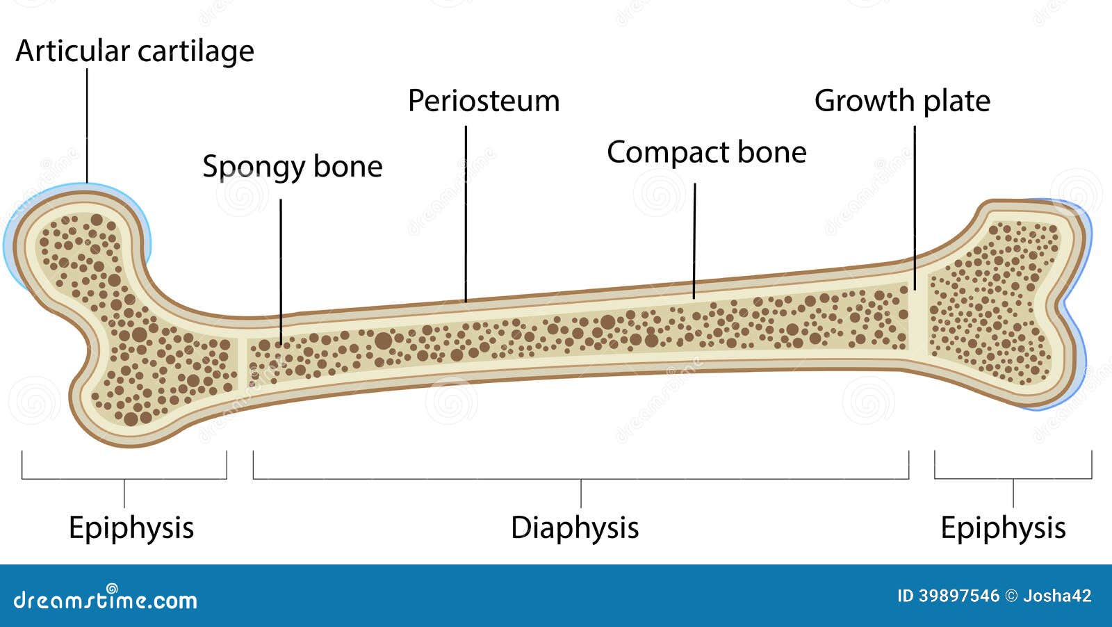
30 Radius And Ulna Diagram - Wire Diagram Source Information from www.purposegames.com 4 lower end presents a tubercle on the posterior surface called as dorsal tubercle of lister. Radius, in anatomy, the outer of the two bones of the forearm when viewed with the palm facing forward. The radius or radial bone is one of the two large bones of the forearm, the other being the ulna. Axial and appendicular system review lab anatomy & physiology 201 with radius anatomy pictures and information these pictures of this page are about:radius bone location. The skeleton acts as a scaffold by providing support and protection for the soft tissues that make up the rest of the body. The radius is a long bone in the forearm. The ulna articulates with the trochlea and the radius articulates with the capitulum. Each bone is a complex living organ that is made up of many cells, protein fibers, and minerals.
The radius bone is shorter.
800 x 701 jpeg 80kb. Be sure to shuffle the deck so that you can really test your self! Long bones are longer than they are wide and are the major bones of the limbs. 1311 chapter 5 upper limb at hendrick. Its upper concave surface articulates with the. The ulna articulates with the trochlea and the radius articulates with the capitulum. It has an upper end, a lower.upper limb. This ulnar view labelled illustration is from 'asklepios atlas of the human anatomy'. I'm not sure of what you mean by bone diagram. 4 lower end presents a tubercle on the posterior surface called as dorsal tubercle of lister. The three joints in the elbow. The radius bone is shorter. The functions of the skeleton are the main difference between radius and ulna is that radius is the long bone that extends from the.
The ulna articulates with the trochlea and the radius articulates with the capitulum labelled radius bone. Close to neck it presents a radial tuberosity.